HARP (a database of Hansen's Disease Antimicrobial Resistance Profiles) is a collection of the predicted structural impacts of missense mutations, with citations to published mutations, in the drug targets of dapsone, rifampicin and ofloxacin, which confer antimicrobial resistance in leprosy. The three drug targets in Mycobacterium leprae, dihydropteroate synthase (DHPS), RNA polymerase (RNAP) and DNA gyrase (GYR), were modelled using homologous protein structures from Mycobacterium tuberculosis. Every amino acid residue in each drug target was mutated to all 19 alternatives, and the impacts of the mutations were predicted with sequence- and structure-based tools and with tools that estimate changes in vibrational entropy.
Vedithi SC, Malhotra S, Skwark MJ, Munir A, Acebrón-García-De-Eulate M, Waman VP, Alsulami A, Ascher DB, Blundell TL. HARP: a database of structural impacts of systematic missense mutations in drug targets of Mycobacterium leprae. Computational and Structural Biotechnology Journal. 2020 Nov 19, doi: https://doi.org/10.1016/j.csbj.2020.11.013
DHPS: The DHPS homodimer was modelled with the multistep protein modelling pipeline (Vivace). The modelled region follows UniProt residue numbering (gene ID ML0224) from 5 to 278 in Chain A. As the DHPS model is a homodimer, mutations can be queried in chain A only (see PMID: 25833404).
RNAP: The heterohexameric RNAP model was built with Vivace (see above), and each chain was modelled as follows (see PMID:32042379)
Chain A -(rpoA -ML1957) – modelled from residue 3 to 226.
Chain B -(rpoA' -ML1957) – modelled from residue 6 to 231.
Chain C -(rpoB -ML1891) – modelled from residue 28 to 1153.
Chain D -(rpoC -ML1890) – modelled from residue 3 to 1281.
Chain E -(rpoZ -ML0542) – modelled from residue 28 to 108.
Chain F -(rpoT -ML1022) – modelled from residue 253 to 574.
GYR: The heterotetrameric model of DNA gyrase (GYR) of Mycobacterium leprae was modelled using PDB ID: 5BS8 as the template. The protein complex (GyrA2GyrB2) is encoded by two genes, gyrA and gyrB.
Chain A -(gyrA -ML0006) – modelled from residue 16 to 501. The intein region at residue positions 131–550 (UniProt numbering) was removed, and residue numbering matches PDB ID 5BS8
Chain B -(gyrB -ML0005) – modelled from residue 440 to 678.
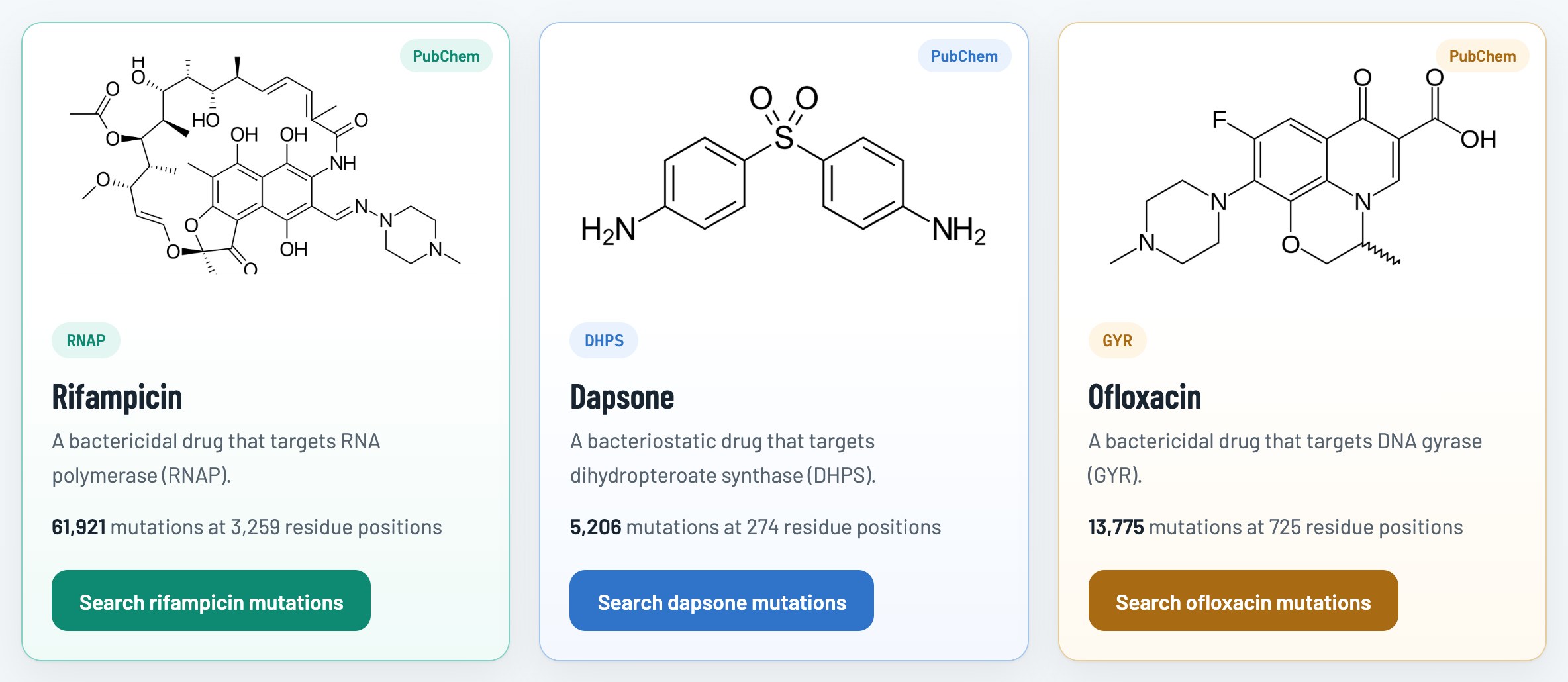
Mutations can be queried from the home page by opening the Mutations menu and choosing a drug:

or by selecting a drug's name, as shown in the figure below:
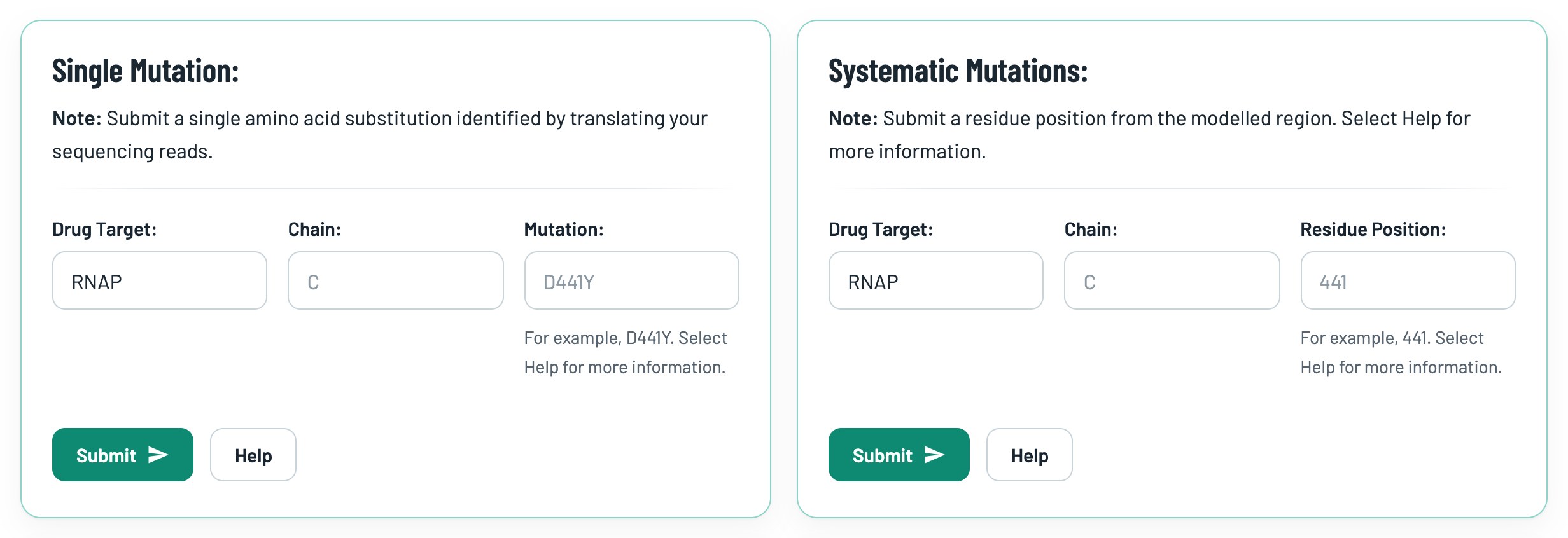
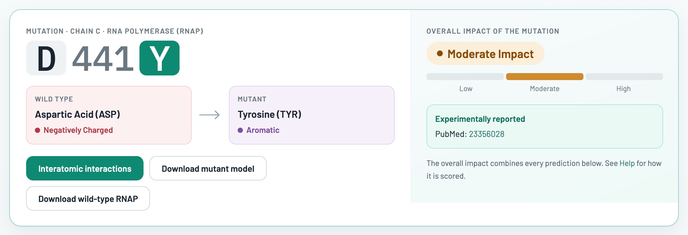
On the Query Mutations page for each drug target, search for a mutation by entering the chain as an uppercase letter (for example, C) and the mutation as the wild-type amino acid (one-letter code), the residue position and the mutant amino acid (one-letter code). For example, D441Y is the substitution of aspartic acid by tyrosine at position 441. Alternatively, enter a residue position in the Systematic Mutations form to see every substitution at that position. To see which regions of each drug target were modelled, see the Modelled Regions section above or select the Help buttons on the Query Mutations page.
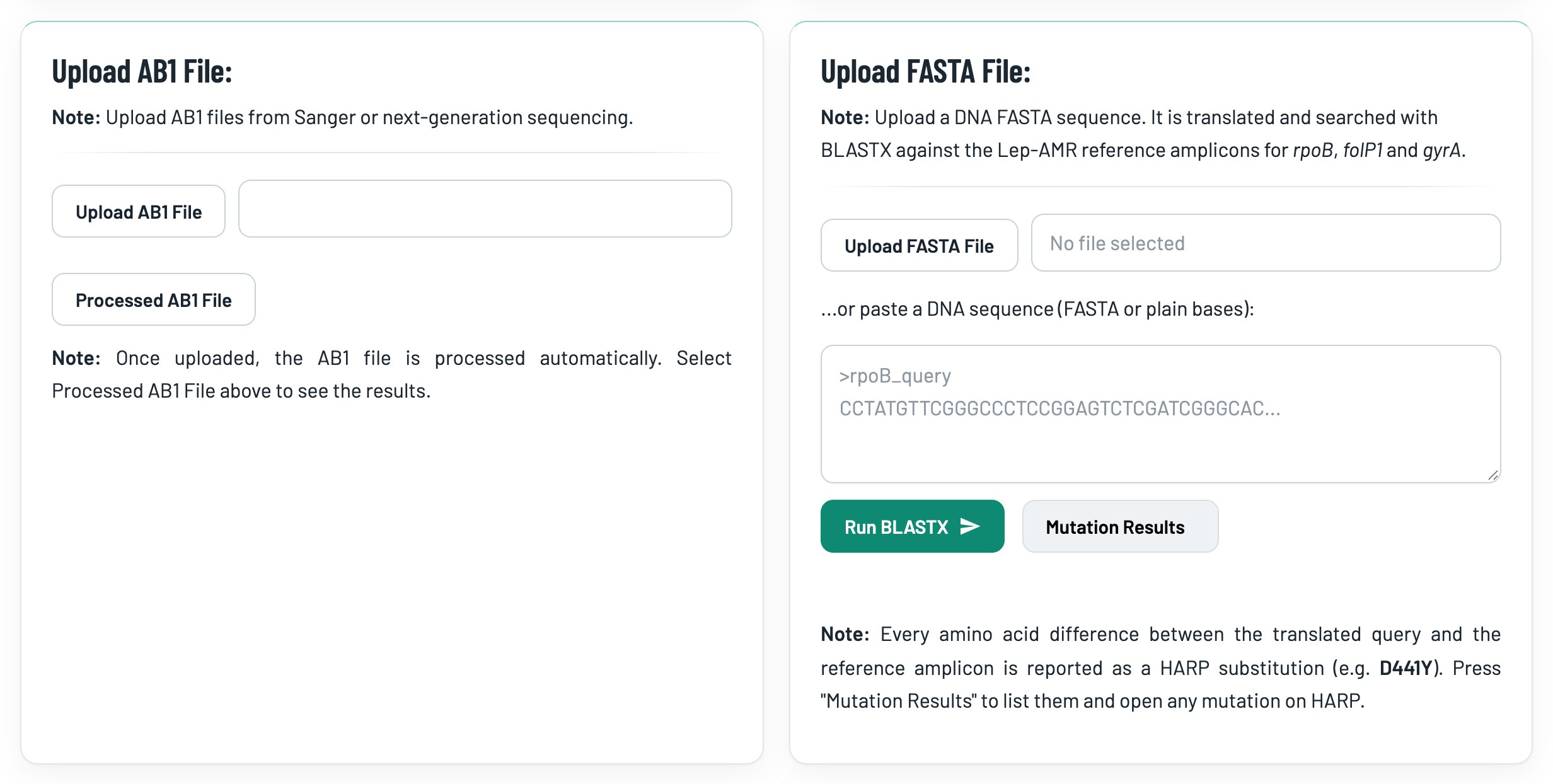
AB1 chromatogram files can be uploaded in the Upload AB1 File panel (figure below). Select the Upload AB1 File button and choose your file. The file uploads immediately and the query page reloads. To view the results, select the Processed AB1 File button.
The results open in a pop-up window (figure below). The top of the window gives the short answer: the gene and residues covered by the trace, and every amino-acid change compared with the Mycobacterium leprae reference, with the overall impact predicted by HARP and a link to the full prediction.
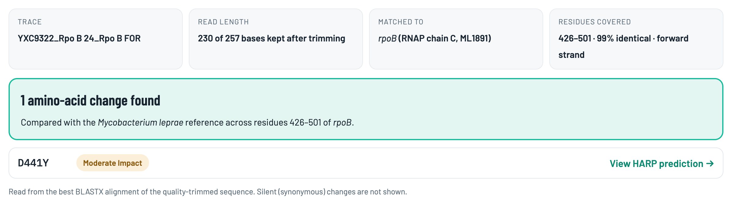
Changes at the very start or end of a read, stop codons and gaps often come from low-quality base calls, so check them in the chromatogram. Silent (synonymous) changes are not shown.
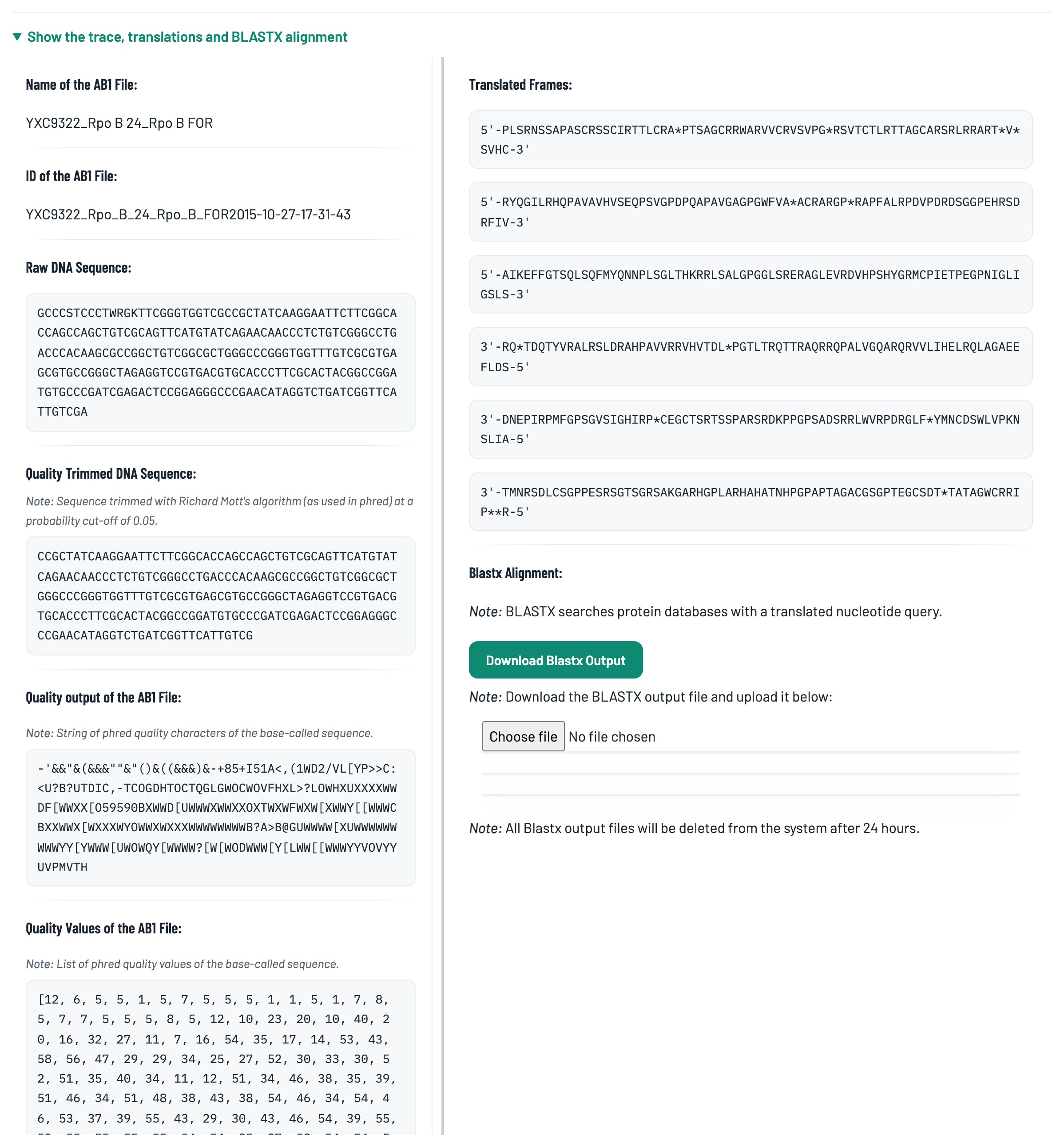
Select Show the trace, translations and BLASTX alignment to see the name and ID of the AB1 file, the raw and quality-trimmed DNA sequences, the quality values, the six translated frames and the BLASTX alignment (figure below). To view the alignment as a graphic, download the BLASTX output with the Download Blastx Output button and upload it with the Choose file button.
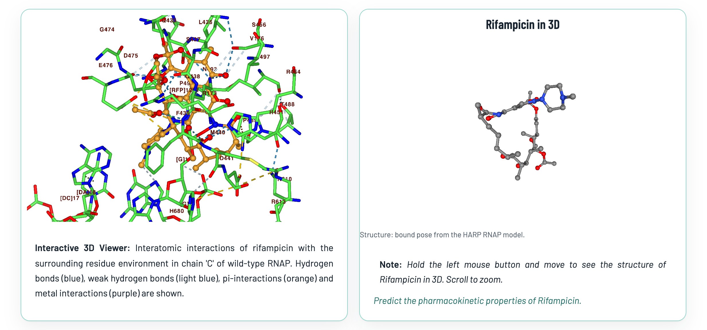
The 3D viewports let you explore the binding site of each drug in its target. Only residues within 5 Å of the ligand are shown in the NGL viewer panel on the left.
The mouse controls for the viewer are as follows:
- Scroll: Zoom scene
- Drag-right: Pan/translate scene
- Drag-left: Rotate scene
- Ctrl-drag right: pan/translate the hovered component
- Ctrl-drag left: rotate the hovered component
- Hover: show a tooltip for the element under the pointer
The right-hand viewport is powered by Molview to view the chemical structure of the ligand.
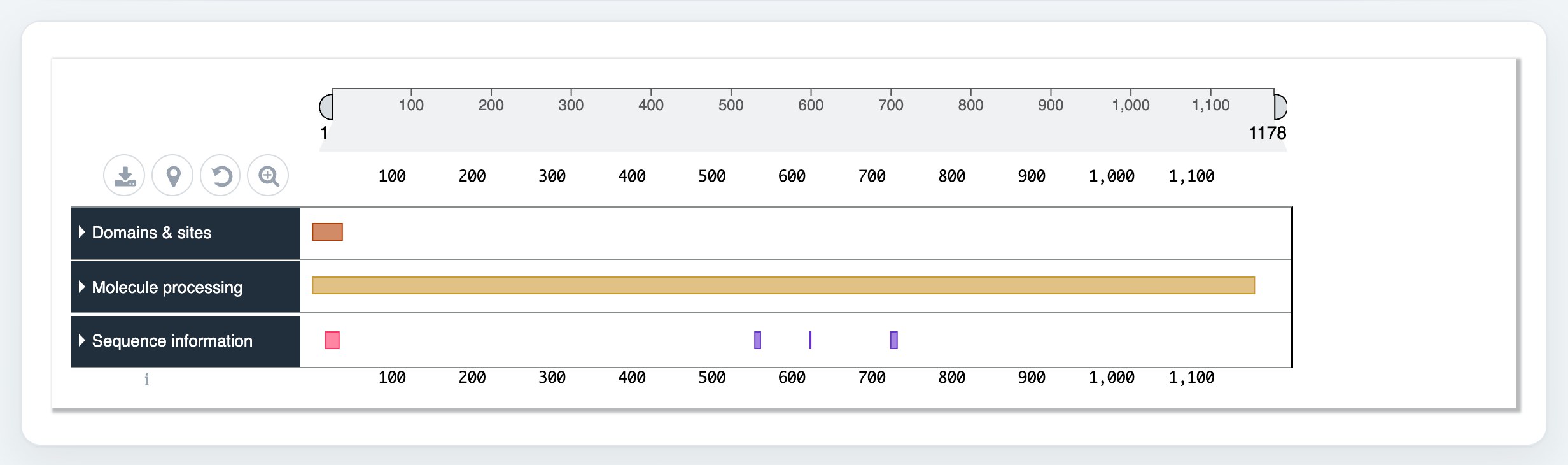
The protein feature viewer presents sequence features in an interactive BioJS component. See the protein feature viewer website for more information. To see the sequence, bring the scale sliders together or scroll over the viewer.
The overall impact of each mutation on protein structure was calculated with a consensus qualitative scoring method, as follows:
I. The qualitative variables are scored as below:
| Property | Outcome |
|---|---|
| Wild-type and mutant residues have the same characteristics (e.g. aliphatic to aliphatic) | Low Impact |
| The mutant residue has different characteristics (e.g. aliphatic to aromatic) | High Impact |
| Conservation score >= 0 (variable residue) (Measured by ConSurf) | Low Impact |
| Conservation score < 0 (conserved residue) | High Impact |
| Interface Residue = No (More than 5Å from the subunit interface) | Low Impact |
| If the mutated residue is an interface residue (less than 5Å from the subunit interface) | High Impact |
| No change in secondary structure due to mutation (identified using SDM2) | Low Impact |
| Change in secondary structure due to mutation | High Impact |
| If the distance of the mutated residue from the ligand is < 5Å | High Impact |
| If the distance of the mutated residue from the ligand is > 5Å | Low Impact |
| If the distance of the mutated residue from the nucleic acid is < 5Å | High Impact |
| If the distance of the mutated residue from the nucleic acid is > 5Å | Low Impact |
II. The following continuous variables are scored using the criteria shown in the figure below: (These values are calculated by SDM2)
- Difference in relative solvent accessibility between the wild type and the mutant residues.
- Difference in depth of the wild-type and mutant residues.
- Difference in occluded packing density of the wild-type and the mutant residues.

II. The stability changes predicted by the following tools are scored using the criteria shown in the figure below:
- mCSM
- mCSM-PPI
- mCSM-NA
- SDM2
- Maestro
- FoldX
- DynaMut

III. For the following tools, the impact of a mutation on protein stability was taken directly from the program output:
- PROVEAN (Neutral, Deleterious)
- CUPSAT (Stabilizing, Neutral, Destabilizing)
- CUPSAT Torsion (Unfavourable, Neutral, Favourable)
- I-Mutant 2.0 (structure) (Decrease, Increase)
- I-Mutant 2.0 (Sequence) (Decrease, Increase)
- EnCOM (Increased Molecular Flexibility, Decreased Molecular Flexibility)
I. Predicted Protein Thermal Stability and Affinity Terms:
The terms for decrease in stability are listed as below (each term has a score of 1):
1. Highly destabilizing
2. Destabilizing
3. Decrease
4. Deleterious
5. Increased Molecular Flexibility
6. Unfavourable
7. Reduced Stability
8. High Impact
9. Moderate Impact
The terms for increase in stability are listed as below (each term has a score of 1):
1. Highly stabilizing
2. Stabilizing
3. Increase
4. Neutral
5. Decreased Molecular Flexibility
6. Favourable
7. Increased Stability
8. Low Impact
9. Neutral
Overall score for each mutation = (Sum of destabilizing terms) - (Sum of stabilizing terms)
The overall score for each mutation is then categorised as shown in the figure below:

* Predictions from the individual tools above are shown on the results pages for single mutations and for systematic mutations (all 19 substitutions).
On the results page for a single mutation, select the Interatomic Interactions button (figure below) to view the wild-type and mutant models:
The molstar viewer is deployed from github repository and customised for HARP. Both the wild-type and the mutant models are loaded by default in the viewer. Select the appropriate model from the icon in the left-hand panel and switch models by clicking the icon. Mol* shows protein structures in a range of representations and displays the interatomic interactions of selected residues. In the figure below, the arrow points to the in-line help icon which gives guidance on using the Mol* viewer. Similar icons in other tabs explain what each tab does.

Interatomic interactions are calculated by Arpeggio, a web-based tool that computes and visualises interatomic interactions in protein structures. You can download the interactions as a comma-separated values (CSV) file.
